

Spots, Freckles & Dullness
Types of Facial Spots and Their Treatments
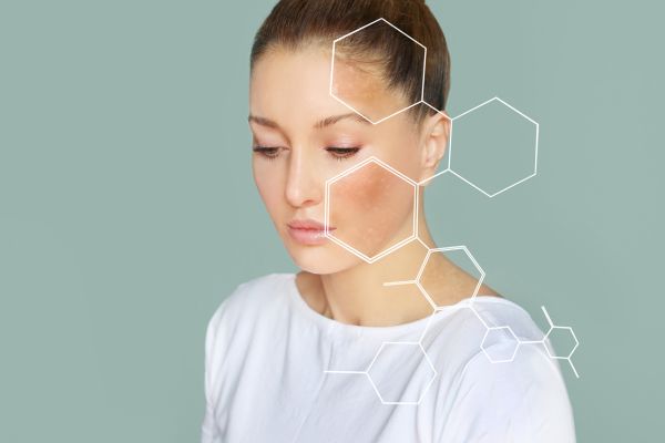
We generally call brown or gray patches on the skin “spots.” But although we lump them together as “spots,” different types have different underlying conditions, so the appropriate treatment differs. Undergoing aesthetic treatment blindly carries a risk of making them worse, so it is important to first receive a correct diagnosis. It is not uncommon for a person to have several of the spots below at once.
Solar Lentigines and Seborrheic Keratosis
A solar lentigo is a spot that appears in almost everyone with age, forming on the face as well as on sun-exposed areas such as the backs of the hands and the arms. It is a brownish spot with a clear border.
When its surface becomes flaky and raised, it is treated as a tumor and called seborrheic keratosis. It is commonly known as a senile wart.
For a solar lentigo, topical hydroquinone or laser treatment is used. With the Discovery Plus we use, the Q-switched YAG laser (wavelength 532 nm), the Q-switched ruby laser (wavelength 694 nm), and the pico YAG laser (wavelength 532 nm) may all be effective, but for those with no particular preference we recommend the pico YAG laser, which is thought to carry a lower risk of post-inflammatory hyperpigmentation.
Learn more aboutDiscovery PICO Plus
For seborrheic keratosis, treatments include liquid nitrogen cryotherapy as well as surgery with a high-frequency electric scalpel and cauterization with a carbon dioxide laser.
Learn more about the high-frequency electric scalpel Learn more about the carbon dioxide laserMelasma
Melasma is a brownish patch seen in women in their 30s to 50s, typically appearing symmetrically high on the cheeks. It can also have a slightly gray tinge.
Unlike a solar lentigo, its border is not distinct. Some doctors even consider melasma to be nothing more than post-inflammatory hyperpigmentation (see below), so in treatment it is most important first to avoid ultraviolet light and to avoid friction.
As oral medications, vitamin C (Cinal) and tranexamic acid (Transamin) are commonly used, and Hythiol (L-cysteine), vitamin E (Juvela), and glutathione (Tathion) may also be used, but the one whose efficacy is most firmly demonstrated by data is tranexamic acid.
Tranexamic acid has the side effect of making blood clots more likely, and vitamin E, being a fat-soluble vitamin, can cause toxic symptoms if taken in excess, so caution is needed.
For topical treatment, hydroquinone, azelaic acid, tretinoin, tranexamic acid, and the like are used. Chemical peeling with glycolic acid and others, or phototherapy (not offered at our clinic), may also be performed.
Also, many people assume that since it is a spot, a laser will work, but applying a laser normally makes melasma worse. (Medically, this is because it is not an organic disease but a functional one.) The technique of applying a laser at a weak setting is commonly called laser toning, but toning is controversial; because it carries a risk of darkening further or causing whitening, it needs to be done at least in combination with other treatments.
There are also various types of laser used for toning; both the Q-switched YAG and the pico YAG laser (fractional and non-fractional) have been reported to be effective, but there are also cases where they are combined with other treatments such as topical hydroquinone.
It has also been pointed out that this data may reflect only the lightening of a different, coexisting spot rather than the melasma itself, and that, because long-term follow-up was not done, recurrence was not captured.
In conclusion, at present our clinic does not recommend laser treatment as a first choice for melasma. When it is performed to treat a different coexisting spot, such as a solar lentigo, care must be taken that the underlying melasma does not worsen.
Ephelides (Freckles)
Ephelides, commonly called freckles, are small brown spots scattered from the cheeks across the nose. They are common in white people and are a hereditary trait. They are present from childhood but fade after puberty, and it is often the case that a coexisting senile spot becomes more conspicuous instead. Laser treatment works well, but because they can recur, care after laser irradiation is also important. With the Discovery Plus we use, the Q-switched YAG laser (wavelength 532 nm), the Q-switched ruby laser (wavelength 694 nm), and the pico YAG laser (wavelength 532 nm) may all be effective, but aiming to remove them in a single session, we recommend the Q-switched ruby laser, which is well absorbed by melanin.
Learn more aboutDiscovery PICO Plus
Nevus of Ota and Acquired Dermal Melanocytosis (ADM)
A nevus of Ota is a blue-gray or brown patch that forms within the first year of life, or by puberty, on one side of the forehead or around the eyes. Pigmented spots can also be seen in the whites of the eyes. Acquired melanosis, on the other hand, is a brown or blue-gray patch that forms after puberty or in adulthood, bilaterally around the eyes and elsewhere. In both, melanocytes, the melanin-producing cells that normally should not be in the dermis, have increased there. Laser treatment is effective, but several sessions are needed. Because post-inflammatory hyperpigmentation occurs after irradiation as a side effect of the laser, rather than rushing to treat it quickly with frequent sessions, it is better to wait at least three months, let it lighten, and perform the next session once the lightening has stopped. At our clinic we can treat it with the Q-switched ruby laser, the Q-switched YAG laser, or the pico YAG laser. For nevus of Ota, the ruby laser and YAG laser are covered by insurance, but the ruby laser is limited to five sessions. Because loss of pigment and the like can occur, irradiation beyond five sessions is not advisable.
Learn more aboutDiscovery PICO Plus
Post-Inflammatory Hyperpigmentation
When some kind of inflammation occurs in the skin, a brown spot can form after the inflammation subsides. On the face, many people are bothered by spots that form after acne, burns, or the eczema of atopic dermatitis, and they are also not uncommon after laser irradiation for treating spots. With atopic dermatitis, it is often not “after the inflammation” but in the very midst of inflammation, so controlling that comes first. In treating post-inflammatory hyperpigmentation, it is important to avoid ultraviolet light, and oral vitamin C or tranexamic acid may be taken. Topical hydroquinone is also good, but continuing to use it while it is causing irritation will make things worse, so caution is needed.
Friction Melanosis
Although it is uncommon, we occasionally encounter it during aesthetic consultations. Pigmentation occurs from rubbing the skin. Its border is indistinct, ranging from a pale brown to a slightly gray tinge. It occurs from rubbing hard while cleansing or from rubbing serum in forcefully. What is in some places called “skincare melasma” is probably, in dermatological terms, this condition. The best treatment is to stop such behavior. Ordinary laser treatment makes it worse. We avoid ultraviolet light and may also use oral vitamin C or tranexamic acid. Topical hydroquinone is also good, but rubbing it in hard, or continuing to use it while it is causing irritation, will make it worse, so caution is needed.
Dullness
When there is no clearly visible spot but the skin somehow looks dull and you want to brighten it by a tone, there may be very faint spots, or spots deep in the skin such as hidden melasma. In addition to oral and topical vitamin C and tranexamic acid, we offer options such as pico toning, ruby fractional, and the Mesona-J brightening course. You can also have a skin analysis with the NeoVoir before treatment.